
Allies story – Part II
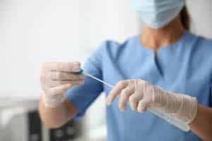
The HPV technician counts the number of leukocytes in the smear test (more than 10-20 in the field of view indicates the presence of infection) and looks for the causative agents of the infection. A smear can reveal fungi (thrush, candidiasis), “key cells” (bacterial vaginosis, gardnerellosis), and changes in the normal flora.
If a smear shows the presence of infection but does not identify the pathogen, the doctor indicates HPV culture and PCR test diagnostics. Treatment of infection must necessarily precede treatment of erosion. Firstly, curing the infection can lead to the spontaneous disappearance of erosion; secondly, surgical treatment of any organ, including the cervix, is impossible in conditions of “contamination” with microbes, leading to complications.
Treatment Approaches
When treating, you must combine general drugs used in tablets and local ones – suppositories, douching, and gels. Local drugs help antibiotics penetrate the affected cells, including erosion cells. Thrush often develops after antibiotic use; to prevent it, the course of antibiotics should end with the use of drugs containing lactic bacteria that normally live in the vagina (for example, Acylact suppositories). The only exception is the initial thrush (candidiasis).
When treating it, acylate is not needed since candidiasis worsens in the presence of a large number of lactic bacteria. Cytological examination is a mandatory stage in diagnosing cervical pathology. Mandatory research was carried out before her treatment. Even if nothing worries you and the cervix seems unchanged to the naked eye, a woman should regularly (every year) undergo a cytological examination of cervical scrapings. Cytology is the science of cells.
Understanding Cervical Changes
The malignant process begins from the cervix’s lower layers of the epithelium (covering tissue) and progresses upward to the surface. Therefore, if the smear only includes the superficial layer, you can make a diagnosis only when the disease has reached its last stage. After scraping cells from a specific area of the cervix—the junction of the integumentary epithelium on the outside and the columnar epithelium lining the cervix canal on the inside—the specialist spreads the cells on a glass slide. The cytologist’s diagnosis may sound like HPV viral load levels are not detected” – normal or “detected”.
Atypical cells are changes in the shape, size, and nuclear structure of cells. A malignant process or inflammation may cause the changes. Therefore, doctors carry out anti-inflammatory treatment in any case and supplement cytology data with a biopsy under colposcopy control. Determining the type of human papillomavirus Sometimes, the doctor suggests conducting a test to determine the type. The fact is that this particular virus causes some types of cervical pathology. And some of its types (16, 18) are highly oncogenic, i.e. are highly likely to cause cervical cancer.
Therefore, with a minor lesion, you can determine the type of virus, remove the highly oncogenic one, and observe the low-oncogenic one. In serious colposcopic pathology, doctors remove the tissue regardless of the virus type, which may not be detected in this case. They determine the virus type using PCR diagnostics.
Colposcopy Procedure
In principle, any gynaecologist must master this test technique so that you can see a colposcope in the office of an ordinary gynaecologist. As a rule, a gynaecologist uses a colposcope to tell whether there is erosion and not to miss pictures suspicious of a malignant tumour. A specialist in cervical pathology performs an extended colposcopy, which involves examining the cervix under a microscope after treating it with special dye solutions containing acetic acid and iodine.
You lie on a gynaecological chair. The HPV doctor inserts a speculum, moves a large device with eyepieces to the chair, turns on the light, and examines the area. This is a colposcope. It remains outside, and its magnification lets you see the cervix afar. The colposcopy procedure is quite long (about 20 minutes); the doctor does it sitting and silently. Therefore, prepare to lie down long and wait to ask the doctor what is there. He’ll be able to answer you accurately only after he’s done a fully extended colposcopy.
Use of Staining Solutions
The first colouring solution used to treat the cervix is a 3% acetic acid solution. It has a specific smell and may cause some tingling, which is completely ok. Acetic acid causes vasospasm, which allows the doctor to examine the cervix itself rather than its vasculature. Before staining with acetic acid, the cervix looks like a large pink spot, after which all existing pathology is visible. The STD doctor pours acetic acid into a speculum from a jar and then blots the cervix with a cotton swab, and looks through the colposcope.
The next solution is the so-called Lugol’s solution. It contains iodine, not an alcohol solution, but an aqueous one. So, Lugol does not sting at all. Lugol’s solution stains normal, healthy cervix cells but does not stain pathologically altered cells. Therefore, this procedure is necessary to see the boundaries of the pathology more clearly. If treatment with solutions causes a strong burning sensation, this is a sign of an inflammatory process. The concentrations of the solutions are not designed for sensations but only for colour. Only inflamed tissue has such increased sensitivity.
Only extended colposcopy can provide an accurate diagnosis of cervical disease and determine its malignancy. Without it, you cannot know the diagnosis. Erosion is an unexplored red spot that can hide anything underneath. The colposcope does not use this word; he makes a diagnosis using generally accepted terms worldwide. The presence of columnar epithelium, normally located inside the canal, on the surface of the cervix. A very common picture in young girls, often present in tests in virgins, becomes complicated by HPV Viral inflammation with the onset of sexual activity. The most common cause is congenital.