
Allies Story – Part III
In girls, the columnar epithelium normally extends beyond the boundaries of the canal. Then he gradually goes inside. This process occurs somewhere before the age of 23-25. Until the entire cylindrical epithelium has gone inside, its remains are visible on the surface – ectopia. If HPV inflammation does not complicate it, this condition is a physiological norm that does not require tests and treatment. Most often, with the onset of sexual activity, microbes settle in the ectopia, preventing it from disappearing on its own and leading to persistent inflammation. In this case, doctors must treat it regardless of age.
Causes of Ectopia
There are other causes of ectopia – hormonal imbalance (ovarian dysfunction), chlamydia, herpes and other infections. Ectropion: A condition that develops after childbirth is an outward inversion of the cervical canal. It looks the same as erosion but occurs after childbirth with a cervix rupture, so scars are visible on the sides of the cervix. Unlike ectopia, it never goes away on its own. It is treated surgically. Leukoplakia: A white plaque that rises above the surface of the cervix – an area of increased keratinisation. It is often a test sign of a chronic HPV lesion infection, most likely viral.
Transformation Zone
Requires biopsy and surgical treatment. Transformation zone (transformation zone) An area of pseudo-erosion healing, altered epithelium, different when stained, especially with Lugol’s solution. It can be typical (normal) and with signs of atypia (keratinised glands, altered blood vessels, irregular keratinisation of the epithelium, condylomas). An atypical transformation zone necessarily requires a biopsy. Condylomas Pointed growths. They occur both on the external genitalia, and on the cervix, and in the vagina. A sign of human papillomavirus infection, the main cause of cervical cancer.
When located on the cervix, a mandatory biopsy and surgical removal are required. Cervical gland cysts In the columnar epithelium of the cervical canal, some glands secrete mucus. With healing ectopia, they remain under the growing normal epithelium, continue producing mucus, and the duct closes. Because of this, they stretch and form cysts. Sometimes, their contents can become infected and fester. Requires opening and removal of contents.
Polyp of the cervical canal Proliferation of columnar epithelium inside the canal. The cause may be infectious or tumour. Requires biopsy and removal. With extensive colposcopy, erosion can be treated because it is unknown what to treat and how dangerous it is. After an extended colposcopy, a specialist in cervical pathology, if necessary, takes a targeted (under the control of a microscope) biopsy from suspicious-looking areas.
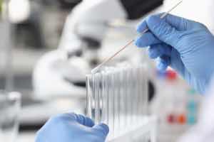
Biopsy
Biopsy: This is the removal of a piece of tissue for histological examination.
Histological HPV examination is the same as cytology, only it examines not individual cells found in a smear, but a whole single section of tissue in a suspicious area. Nuances:
– The doctor performs the biopsy at the beginning of the menstrual cycle, on days 5-7 of the cycle (where day 1 is the first day of menstruation), immediately after menstruation ends.
– The doctor performs the biopsy only in the absence of infection.
If the results of smears for flora are poor, the doctor prescribes treatment for the infection and then takes a biopsy after a good control smear.
Procedure Details
Because a biopsy is a small operation that requires cleanliness, doctors perform it under the control of a colposcope and use Lugol’s solution to visualize the changed area. They use special biopsy forceps, a radio knife (Surgitron device), an electric loop, or a scalpel to perform the biopsy. The procedure is very quick and painless. You take a deep breath at the doctor’s command, and they take the biopsy. Therefore, doctors perform this procedure without pain relief. But if you insist, the doctor may spray a lidocaine solution on the cervix.
The secret is that there are no pain endings on the cervix, so lidocaine has no effect at all; it is a psychological measure against capricious patients. You will not feel pain; discomfort in the lower abdomen (straining) occurs because the uterus contracts in response to touching the cervix. You can eliminate this reaction only by relaxing. By tensing, you increase muscle contraction. – The biopsy site may bleed. The doctor may cauterise this point and press it with a gauze pad.
The cauterisation lasts a few seconds and causes a tugging sensation in the lower abdomen, as during menstruation. – for a week after the biopsy, you need to take care of the cervix – do not be sexually active, do not douche, do not lift heavy objects. The histological examination allows us to make a final diagnosis.
Histological Examination Results
Histological examination may show the following conditions: Chronic cervicitis.
This diagnosis does not require urgent surgical treatment. Instead, anti-inflammatory and possibly antiviral treatment is indicated. Additionally, squamous metaplasia is a natural, normal process of healing “erosion” by replacing the normal columnar epithelium of the cervix with a flat one. The metaplastic epithelium, which appears in the colposcopic picture of the transformation zone, is normal and does not require any treatment.
On the other hand, HPV and leukoplakia, along with acanthosis, dyskeratosis, and hyperkeratosis, involve disorders of epithelial maturation and keratinization; thus, surgical testing and treatment are indicated. Lastly, flat condyloma requires evaluation and management based on its specific characteristics.
Signs of Leukoplakia and Koilocytosis
Signs of leukoplakia and koilocytosis (viral cell damage) indicate that a chronic viral infection causes dyskeratosis. HPV Doctors prescribe antiviral tests and treatment, repeat colposcopy and biopsy, and consider surgical treatment. The issue is that changes in tissue do not always show up during colposcopy. In flat condyloma, dysplasia can develop, and externally, it will remain unchanged. In addition, not all of the affected tissue is included in the biopsy site. If flat condyloma does not respond to antiviral treatment, it is worth considering surgery. Dysplasia. Violation of the structure of the cervical epithelium.
There are three degrees of severity. Mild and moderate (grade 1-2) dysplasia can be observed for some time. Severe dysplasia (grade 3) is an indication for surgical treatment. Dysplasia is a benign change, not always of a tumour nature; it often has an inflammatory cause. Therefore, anti-inflammatory, antiviral treatment and a repeat biopsy are first necessary for detecting dysplasia. The degree of dysplasia after anti-inflammatory treatment will already be true tumour dysplasia, carcinoma in situ, and cervical cancer.
These diagnoses require treatment by an oncologist, not a gynaecologist. Based on the histological examination results, the test doctor chooses the HPV treatment method for cervical erosion. Doctors should use medications only to treat inflammation.